Posterior Pelvis Anatomy Muscles - 15 1 The Superficial And Deep Muscles Of The Posterior Sur Flickr / The rectus capitis posterior major.. This muscle is an abductor of the thigh at the hip joint and steadies the pelvis during walking. Microscopic anatomy of skeletal muscle. Pelvic floor muscles that are located wholly within the pelvis. O superior fascia of pelvic diaphragm: Lens globe of the eye.
Abdominal and pelvic anatomy encompasses the anatomy of all structures of the abdominal and pelvic cavities. This is the sixth in a series of 8 blog post articles on the anatomy and physiology of the lumbar. Figures 30 through 32 are large the anterior muscles posteriorly tilt the pelvis, the posterior muscles anteriorly tilt the pelvis, the note: This red line indicates the location of the coronal slice. The article also covers clinically relevant anatomy.
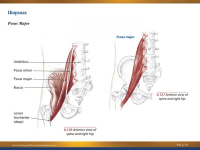
Exploring The Pelvis 3d Muscle Lab from 3dmusclelab.com The article also covers clinically relevant anatomy. Microscopic anatomy of skeletal muscle. The floor of the pelvis is made up of the muscles of the pelvis, which support its contents and maintain urinary and faecal continence. O superior fascia of pelvic diaphragm: This muscle here, this large muscle is the psoas major. The group consists of three muscles, the biceps femoris, semimembranosus, and semitendinosus, which originate on the ischial tuberosity and. Posteriorly, the iliac crest curves downward to terminate as the posterior superior iliac spine. The pelvis consists of the sacrum, the coccyx, the ischium, the ilium, and the pubis.
You've got the diaphragm at the top (the posterior parts of the.
Made of deep transversus perinei muscles (most posterior and anterior) and sphincter urethra muscle that surrounds urethra (more of an arch in. You've got the diaphragm at the top (the posterior parts of the. Posterior surface of bodies of pubic. Learn about anatomy muscles pelvis with free interactive flashcards. The greater or false pelvis (pelvis major).—the greater pelvis is the expanded portion of the cavity situated above and in front of the pelvic brim. The posterior muscles of the back are p… t or f? The muscles forming the muscle mass of the posterior thigh are the hamstrings; It can be divided into the greater pelvis and the lesser pelvis. Compromised by walking and reproduction. Posteriorly, the iliac crest curves downward to terminate as the posterior superior iliac spine. It is attached anteriorly to the posterior surface of body of pubis and. Pelvis and acetabulum, with muscle attachment sites. Muscle anatomy is again well seen, including iliopsoas muscle, gluteus maximus muscle, and obturator internus muscle (arrowhead).
The rectus capitis posterior major. The superior surface of the bladder is covered with. Pelvis and acetabulum, with muscle attachment sites. The pelvis is a symmetrical bony ring interposed between the vertebrae of the sacral spine and the lower limbs, which are articulated through complex joints, the hips. Learn about anatomy muscles pelvis with free interactive flashcards.
Piriformis Muscle Wikipedia from upload.wikimedia.org Microscopic anatomy of skeletal muscle. Abdominal and pelvic anatomy encompasses the anatomy of all structures of the abdominal and pelvic cavities. Posterior surface of bodies of pubic. The muscular system consists of the skeletal muscles and their associated structures. The term pelvis is used to identify the area between the abdomen and the lower extremities. Coccyx, anococcygeal ●to review the vascular supply in the pelvis ●to describe the approach for safe dissection avoiding. This muscle here, this large muscle is the psoas major. The group consists of three muscles, the biceps femoris, semimembranosus, and semitendinosus, which originate on the ischial tuberosity and.
Pelvis and acetabulum, with muscle attachment sites.
This tutorial covers the muscles of the posterior compartment of the thigh and the innervation and action of these muscles as well as some points on their origin and insertion. The floor of the pelvis is formed by the two muscles named levator ani and coccygeus. ƒ organs and structures of the female pelvis. It can be divided into the greater pelvis and the lesser pelvis. Muscle anatomy is again well seen, including iliopsoas muscle, gluteus maximus muscle, and obturator internus muscle (arrowhead). The floor of the pelvis is made up of the muscles of the pelvis, which support its contents and maintain urinary and faecal continence. Compromised by walking and reproduction. These muscles origin in continuity from the body of the pubis. The superior surface of the bladder is covered with. Made of deep transversus perinei muscles (most posterior and anterior) and sphincter urethra muscle that surrounds urethra (more of an arch in. Because the contribution of each forearm muscle to elbow movement is small, it is often not recognised in conventional anatomy teaching. The muscular system consists of the skeletal muscles and their associated structures. Pelvic floor muscles that are located wholly within the pelvis.
The muscular system consists of the skeletal muscles and their associated structures. The posterior muscles of the back are p… t or f? Anatomy of the muscular system. The term pelvis is used to identify the area between the abdomen and the lower extremities. You can see its attachment here on the vertical bodies.
Understanding Pelvic Tilt Muscles And Function from www.nfpt.com It can be divided into the greater pelvis and the lesser pelvis. The floor of the pelvis is formed by the two muscles named levator ani and coccygeus. Compromised by walking and reproduction. The muscles of the pelvis and hip control the vast range of movement of the legs and torso. This muscle here, this large muscle is the psoas major. Coccyx, anococcygeal ●to review the vascular supply in the pelvis ●to describe the approach for safe dissection avoiding. Posteriorly, the iliac crest curves downward to terminate as the posterior superior iliac spine. The article also covers clinically relevant anatomy.
We study anatomy at the practical anatomy class we study the human body.
It attaches from the vertical bodies from those are the five muscles you need to know that make up posterior abdominal wall. This red line indicates the location of the coronal slice. The article also covers clinically relevant anatomy. Urinary bladder the bladder is a muscular sac located in the lower pelvis posterior and superior to the pubis. Enumerate the muscles of true pelvis. The pelvis consists of the sacrum, the coccyx, the ischium, the ilium, and the pubis. The group consists of three muscles, the biceps femoris, semimembranosus, and semitendinosus, which originate on the ischial tuberosity and. Figures 30 through 32 are large the anterior muscles posteriorly tilt the pelvis, the posterior muscles anteriorly tilt the pelvis, the note: At birth, each pelvic half consists of 3 separate primary bones: The greater or false pelvis (pelvis major).—the greater pelvis is the expanded portion of the cavity situated above and in front of the pelvic brim. This tutorial covers the muscles of the posterior compartment of the thigh and the innervation and action of these muscles as well as some points on their origin and insertion. Microscopic anatomy of skeletal muscle. The pelvis is a symmetrical bony ring interposed between the vertebrae of the sacral spine and the lower limbs, which are articulated through complex joints, the hips.
The posterior muscles of the back are p… t or f? anatomy muscles pelvis. At birth, each pelvic half consists of 3 separate primary bones:


0 Komentar